
1
Introduction
1 min•161 words
Traumatic Brain Injury stands as one of the most devastating global public health crises, representing a leading cause of permanent neurological disability, economic hardship, and premature mortality. The burden of neuro-trauma is disproportionately high in low- and middle-income countries, particularly within sub-Saharan Africa, where developing emergency response systems and resource-constrained tertiary centers struggle to cope with an influx of complex cases.
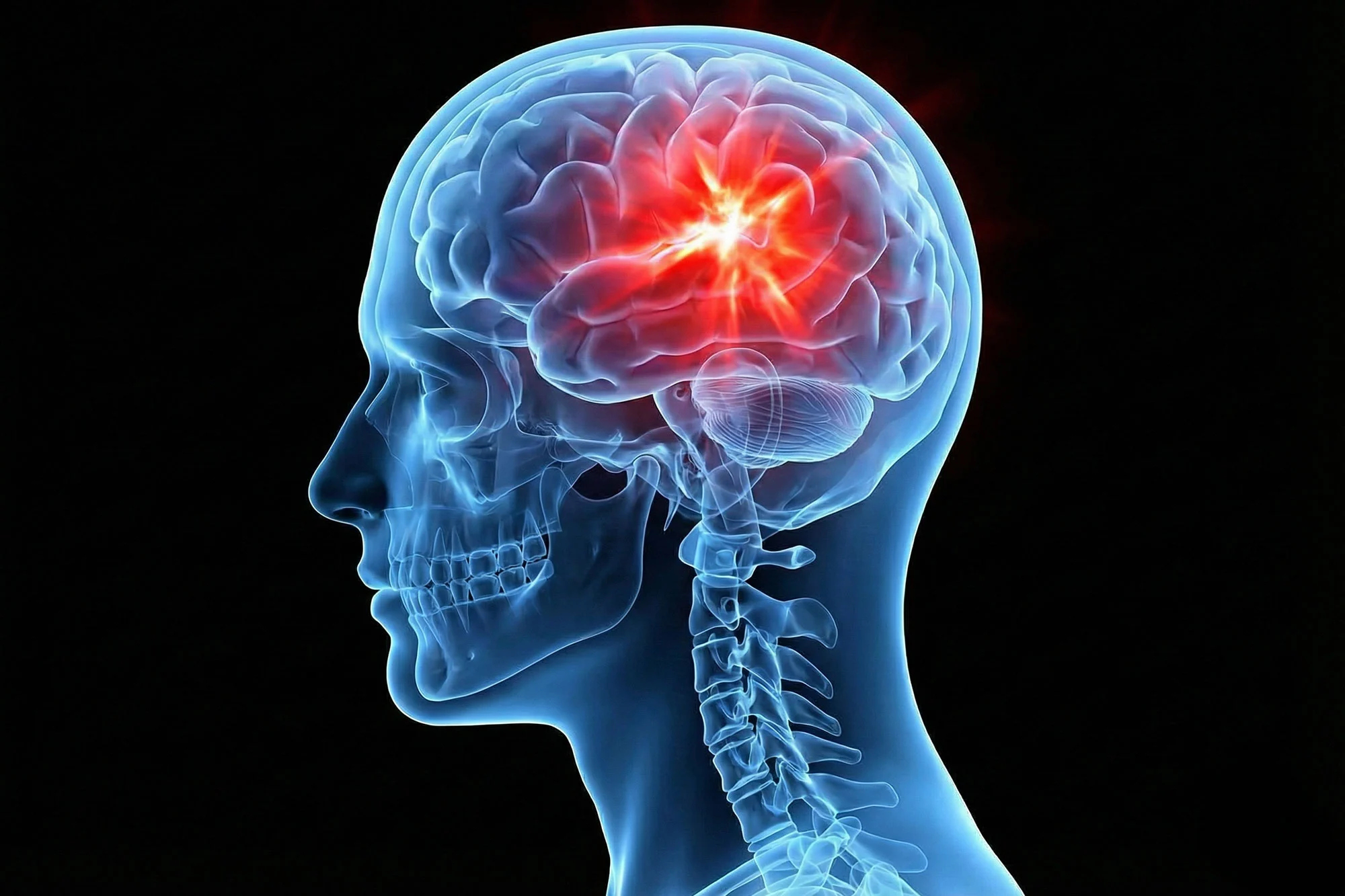
Clinically, a head injury is defined as any structural alteration or trauma to the scalp, skull, or brain tissue caused by an external mechanical force, explicitly excluding superficial injuries confined to the face. When this mechanical energy disrupts normal brain function temporarily or permanently, it is designated as a Traumatic Brain Injury.
In settings like Uganda, neuro-trauma is concentrated heavily at national referral centers. For instance, data from Mulago National Referral Hospital reveals an alarming cumulative incidence of head injuries, demonstrating that without early, protocolized emergency interventions, these injuries rapidly progress from initial structural disruptions to irreversible secondary brain damage.
Clinically, a head injury is defined as any structural alteration or trauma to the scalp, skull, or brain tissue caused by an external mechanical force, explicitly excluding superficial injuries confined to the face. When this mechanical energy disrupts normal brain function temporarily or permanently, it is designated as a Traumatic Brain Injury.
In settings like Uganda, neuro-trauma is concentrated heavily at national referral centers. For instance, data from Mulago National Referral Hospital reveals an alarming cumulative incidence of head injuries, demonstrating that without early, protocolized emergency interventions, these injuries rapidly progress from initial structural disruptions to irreversible secondary brain damage.
2
Background & Pathophysiology
2 min•248 words
The pathophysiology of Traumatic Brain Injury is fundamentally split into two distinct processes: primary tissue injury and secondary tissue injury.
Primary brain injury occurs at the exact microsecond of impact. It is the direct result of mechanical forces causing immediate macroscopic and microscopic damage to the intracranial structures. This includes focal contusions, lacerations of the brain parenchyma, skull fractures, tearing of blood vessels leading to epidural, subdural, or intracerebral hematomas, and diffuse axonal injury caused by acceleration-deceleration shearing forces. This primary damage is irreversible at the bedside and can only be mitigated through population-level preventative strategies like road safety enforcement.
Secondary brain injury, conversely, is a progressive cascade of neurobiological destruction that develops minutes, hours, and days following the initial impact. The injured, vulnerable brain tissue suffers from a massive release of excitatory neurotransmitters like glutamate, leading to intracellular calcium overload, mitochondrial dysfunction, free radical production, and widespread neuro-inflammation. This cascade triggers severe cerebral edema, which compromises local microvascular blood flow.
Because the skull is a rigid, unyielding container, worsening cerebral edema or expanding hematomas cause a dangerous rise in intracranial pressure. If intracranial pressure exceeds the systemic blood pressure, the cerebral perfusion pressure drops, plunging the brain tissue into profound ischemia. This ischemic penumbra is highly vulnerable to systemic insults. The primary goal of acute medical management is to aggressively prevent and eliminate the "deadly twins" of secondary brain injury: systemic hypotension (which drops cerebral perfusion) and systemic hypoxia (which starves the ischemic brain tissue of oxygen).
Primary brain injury occurs at the exact microsecond of impact. It is the direct result of mechanical forces causing immediate macroscopic and microscopic damage to the intracranial structures. This includes focal contusions, lacerations of the brain parenchyma, skull fractures, tearing of blood vessels leading to epidural, subdural, or intracerebral hematomas, and diffuse axonal injury caused by acceleration-deceleration shearing forces. This primary damage is irreversible at the bedside and can only be mitigated through population-level preventative strategies like road safety enforcement.
Secondary brain injury, conversely, is a progressive cascade of neurobiological destruction that develops minutes, hours, and days following the initial impact. The injured, vulnerable brain tissue suffers from a massive release of excitatory neurotransmitters like glutamate, leading to intracellular calcium overload, mitochondrial dysfunction, free radical production, and widespread neuro-inflammation. This cascade triggers severe cerebral edema, which compromises local microvascular blood flow.
Because the skull is a rigid, unyielding container, worsening cerebral edema or expanding hematomas cause a dangerous rise in intracranial pressure. If intracranial pressure exceeds the systemic blood pressure, the cerebral perfusion pressure drops, plunging the brain tissue into profound ischemia. This ischemic penumbra is highly vulnerable to systemic insults. The primary goal of acute medical management is to aggressively prevent and eliminate the "deadly twins" of secondary brain injury: systemic hypotension (which drops cerebral perfusion) and systemic hypoxia (which starves the ischemic brain tissue of oxygen).
3
Clinical Features
2 min•285 words
The clinical presentation of Traumatic Brain Injury varies drastically depending on the anatomical location of the trauma, the mass effect of expanding intracranial lesions, and the baseline physiological reserve of the patient.
History taking must occur concurrently with stabilization and focus heavily on the mechanism of injury, the presence or absence of a witnessed loss of consciousness, post-traumatic amnesia (both anterograde and retrograde), episodes of projectile vomiting, and any history of post-traumatic seizures.
Physical examination relies heavily on objective clinical scoring systems. Neurological responsiveness is initially screened using the rapid AVPU scale, which classifies patients as Alert, responding to Voice, responding only to Pain, or completely Unresponsive. For comprehensive monitoring, the Glasgow Coma Scale is the gold standard, evaluating eye opening, verbal response, and motor responsiveness.
Based on the Glasgow Coma Scale, head injuries are strictly stratified into three categories of severity:
Mild Traumatic Brain Injury is defined by a Glasgow Coma Scale score of 13 to 15. These patients are conscious but may complain of transient confusion, headaches, dizziness, or short-term memory gaps.
Moderate Traumatic Brain Injury is defined by a Glasgow Coma Scale score of 9 to 12. These individuals frequently present with significant lethargy, confusion, or focal neurological deficits.
Severe Traumatic Brain Injury is defined by a Glasgow Coma Scale score of 3 to 8, representing a comatose patient who is entirely incapable of protecting their own airway.
Beyond the Glasgow Coma Scale, the clinician must examine the head carefully for physical signs of trauma. This includes inspecting for open or depressed skull deformities, and checking for pathognomonic signs of a basal skull fracture, which include raccoon eyes (bilateral periorbital ecchymosis), Battle's sign (post-auricular ecchymosis), hemotympanum, and cerebrospinal fluid rhinorrhea or otorrhea.
History taking must occur concurrently with stabilization and focus heavily on the mechanism of injury, the presence or absence of a witnessed loss of consciousness, post-traumatic amnesia (both anterograde and retrograde), episodes of projectile vomiting, and any history of post-traumatic seizures.
Physical examination relies heavily on objective clinical scoring systems. Neurological responsiveness is initially screened using the rapid AVPU scale, which classifies patients as Alert, responding to Voice, responding only to Pain, or completely Unresponsive. For comprehensive monitoring, the Glasgow Coma Scale is the gold standard, evaluating eye opening, verbal response, and motor responsiveness.
Based on the Glasgow Coma Scale, head injuries are strictly stratified into three categories of severity:
Mild Traumatic Brain Injury is defined by a Glasgow Coma Scale score of 13 to 15. These patients are conscious but may complain of transient confusion, headaches, dizziness, or short-term memory gaps.
Moderate Traumatic Brain Injury is defined by a Glasgow Coma Scale score of 9 to 12. These individuals frequently present with significant lethargy, confusion, or focal neurological deficits.
Severe Traumatic Brain Injury is defined by a Glasgow Coma Scale score of 3 to 8, representing a comatose patient who is entirely incapable of protecting their own airway.
Beyond the Glasgow Coma Scale, the clinician must examine the head carefully for physical signs of trauma. This includes inspecting for open or depressed skull deformities, and checking for pathognomonic signs of a basal skull fracture, which include raccoon eyes (bilateral periorbital ecchymosis), Battle's sign (post-auricular ecchymosis), hemotympanum, and cerebrospinal fluid rhinorrhea or otorrhea.
4
Diagnosis & Workup
2 min•344 words
In resource-limited settings, the diagnostic workup must be highly focused to avoid delaying life-saving resuscitations and to ensure expensive imaging modalities are utilized efficiently.
Triage and Clinical Screening: Every patient presenting with a head injury must undergo immediate triage. Red flag triggers that demand immediate medical officer activation within thirty minutes of arrival include any signs of airway obstruction, a respiratory rate below ten or above twenty-nine breaths per minute, a systolic blood pressure below ninety mmHg, or an AVPU score worse than Alert. To evaluate poly-trauma patients holistically, general trauma scoring tools like the Kampala Trauma Score should be computed alongside the Glasgow Coma Scale.
Protocolized Adult CT Scan Criteria: A non-contrast computed tomography scan of the head is the definitive diagnostic modality for structural intracranial pathology. Due to cost and availability constraints, a head CT must be ordered strictly if the adult patient meets any of the following high-risk criteria: a Glasgow Coma Scale score below thirteen on initial evaluation or below fifteen at two hours post-injury; any suspected open, depressed, or basal skull fracture; post-traumatic seizures; focal neurological deficits; more than one episode of vomiting; retrograde amnesia exceeding thirty minutes; age sixty-five years or older; known bleeding disorders or active anticoagulant use; or a high-risk mechanism of injury such as being a pedestrian struck by a vehicle, ejection from a vehicle, or falling from a height exceeding three feet.
Protocolized Pediatric CT Scan Criteria: Because the developing pediatric brain is highly sensitive to radiation, CT imaging in children under sixteen years must be heavily rationalized. A head CT is indicated immediately if a child exhibits a witnessed loss of consciousness lasting more than five minutes; anterograde or retrograde amnesia lasting more than five minutes; abnormal, persistent drowsiness; three or more distinct episodes of vomiting; clinical suspicion of non-accidental injury; a post-traumatic seizure without a history of epilepsy; a GCS below fourteen (or pediatric GCS below fifteen for infants under one year); a tense fontanelle; or any head bruise, swelling, or laceration exceeding five centimeters in an infant under twelve months old.
Triage and Clinical Screening: Every patient presenting with a head injury must undergo immediate triage. Red flag triggers that demand immediate medical officer activation within thirty minutes of arrival include any signs of airway obstruction, a respiratory rate below ten or above twenty-nine breaths per minute, a systolic blood pressure below ninety mmHg, or an AVPU score worse than Alert. To evaluate poly-trauma patients holistically, general trauma scoring tools like the Kampala Trauma Score should be computed alongside the Glasgow Coma Scale.
Protocolized Adult CT Scan Criteria: A non-contrast computed tomography scan of the head is the definitive diagnostic modality for structural intracranial pathology. Due to cost and availability constraints, a head CT must be ordered strictly if the adult patient meets any of the following high-risk criteria: a Glasgow Coma Scale score below thirteen on initial evaluation or below fifteen at two hours post-injury; any suspected open, depressed, or basal skull fracture; post-traumatic seizures; focal neurological deficits; more than one episode of vomiting; retrograde amnesia exceeding thirty minutes; age sixty-five years or older; known bleeding disorders or active anticoagulant use; or a high-risk mechanism of injury such as being a pedestrian struck by a vehicle, ejection from a vehicle, or falling from a height exceeding three feet.
Protocolized Pediatric CT Scan Criteria: Because the developing pediatric brain is highly sensitive to radiation, CT imaging in children under sixteen years must be heavily rationalized. A head CT is indicated immediately if a child exhibits a witnessed loss of consciousness lasting more than five minutes; anterograde or retrograde amnesia lasting more than five minutes; abnormal, persistent drowsiness; three or more distinct episodes of vomiting; clinical suspicion of non-accidental injury; a post-traumatic seizure without a history of epilepsy; a GCS below fourteen (or pediatric GCS below fifteen for infants under one year); a tense fontanelle; or any head bruise, swelling, or laceration exceeding five centimeters in an infant under twelve months old.
5
Management
3 min•426 words
The management of Traumatic Brain Injury demands a strict, timeline-driven protocol that prioritizes systemic resuscitation to shield the brain from secondary ischemic degradation.
Airway and Oxygenation Optimization: Ensuring adequate oxygenation is a cornerstone of therapy. Administer immediate, continuous high-flow supplemental oxygen via a non-rebreather mask to all patients with severe Traumatic Brain Injury, keeping target oxygen saturations strictly above ninety percent. For patients with a Glasgow Coma Scale score of eight or less, the airway is structurally unsafe. Advanced airway management, including endotracheal intubation or a definitive surgical airway, must be performed early by trained personnel. If intensive care infrastructure is unavailable to support an intubated patient, this must be immediately documented, the family counseled, and senior surgical leadership contacted.
Hemodynamic Fluid Resuscitation: Systemic hypotension must be corrected aggressively, as a single episode of a systolic blood pressure below ninety mmHg doubles the mortality rate in severe head injuries. Fluid resuscitation must be achieved using isotonic crystalloids, specifically Normal Saline or Ringer's Lactate, to maintain a target systolic blood pressure above ninety mmHg. It is an absolute, critical contraindication to use five percent Dextrose or alternative hypotonic fluids in head injury management, as these fluids rapidly drive water into the injured brain tissue, causing catastrophic cerebral edema and fatal intracranial herniation.
Protocolized Pain and Seizure Management: Pain must be actively managed using objective pain scales. First-line therapy includes intravenous Paracetamol at ten to fifteen milligrams per kilogram. Second-line treatment for adults and older adolescents includes intravenous Tramadol. However, Tramadol is strictly contraindicated in pediatric patients under twelve years of age. For severe pain, intravenous Fentanyl or Morphine may be used under continuous respiratory monitoring. To prevent further secondary hypoxia from convulsions, any post-traumatic seizure must be treated immediately with an intravenous loading dose of Phenytoin at fifteen to twenty milligrams per kilogram, or Phenobarbitone if Phenytoin is unavailable or contraindicated.
Targeted Hyperosmolar and Metabolic Control: Hyperosmolar therapy using intravenous Mannitol at a dose of zero point twenty-five to one gram per kilogram is highly effective at reducing elevated intracranial pressure by drawing fluid out of the brain parenchyma. However, Mannitol is an osmotic diuretic that causes severe systemic hypotension. Therefore, Mannitol is strictly contraindicated in any patient with a systolic blood pressure below ninety mmHg. If a hypotensive patient shows signs of intracranial herniation, hypertonic saline must be used instead of Mannitol. Additionally, corticosteroids are completely unrecommended and must not be administered for traumatic brain injury. Prior to discharge or transfer from the emergency department, every patient with an open injury must receive standard Tetanus prophylaxis.
Airway and Oxygenation Optimization: Ensuring adequate oxygenation is a cornerstone of therapy. Administer immediate, continuous high-flow supplemental oxygen via a non-rebreather mask to all patients with severe Traumatic Brain Injury, keeping target oxygen saturations strictly above ninety percent. For patients with a Glasgow Coma Scale score of eight or less, the airway is structurally unsafe. Advanced airway management, including endotracheal intubation or a definitive surgical airway, must be performed early by trained personnel. If intensive care infrastructure is unavailable to support an intubated patient, this must be immediately documented, the family counseled, and senior surgical leadership contacted.
Hemodynamic Fluid Resuscitation: Systemic hypotension must be corrected aggressively, as a single episode of a systolic blood pressure below ninety mmHg doubles the mortality rate in severe head injuries. Fluid resuscitation must be achieved using isotonic crystalloids, specifically Normal Saline or Ringer's Lactate, to maintain a target systolic blood pressure above ninety mmHg. It is an absolute, critical contraindication to use five percent Dextrose or alternative hypotonic fluids in head injury management, as these fluids rapidly drive water into the injured brain tissue, causing catastrophic cerebral edema and fatal intracranial herniation.
Protocolized Pain and Seizure Management: Pain must be actively managed using objective pain scales. First-line therapy includes intravenous Paracetamol at ten to fifteen milligrams per kilogram. Second-line treatment for adults and older adolescents includes intravenous Tramadol. However, Tramadol is strictly contraindicated in pediatric patients under twelve years of age. For severe pain, intravenous Fentanyl or Morphine may be used under continuous respiratory monitoring. To prevent further secondary hypoxia from convulsions, any post-traumatic seizure must be treated immediately with an intravenous loading dose of Phenytoin at fifteen to twenty milligrams per kilogram, or Phenobarbitone if Phenytoin is unavailable or contraindicated.
Targeted Hyperosmolar and Metabolic Control: Hyperosmolar therapy using intravenous Mannitol at a dose of zero point twenty-five to one gram per kilogram is highly effective at reducing elevated intracranial pressure by drawing fluid out of the brain parenchyma. However, Mannitol is an osmotic diuretic that causes severe systemic hypotension. Therefore, Mannitol is strictly contraindicated in any patient with a systolic blood pressure below ninety mmHg. If a hypotensive patient shows signs of intracranial herniation, hypertonic saline must be used instead of Mannitol. Additionally, corticosteroids are completely unrecommended and must not be administered for traumatic brain injury. Prior to discharge or transfer from the emergency department, every patient with an open injury must receive standard Tetanus prophylaxis.
6
Key Pearls & Takeaways
1 min•191 words
rotect the Penumbra: Primary brain injury cannot be reversed, but secondary brain injury can be prevented. Your primary clinical mandate is to eliminate hypoxia and hypotension, the two most lethal drivers of secondary brain damage.
The Dextrose Danger: Never administer five percent Dextrose fluids to a patient with a traumatic brain injury. Hypotonic fluids worsen cerebral edema, increase intracranial pressure, and can directly precipitate brain herniation.
Know Your Mannitol Limits: Never administer Mannitol to a patient who is hypotensive with a systolic blood pressure below ninety mmHg. Mannitol's diuretic effect will worsen circulatory shock; use hypertonic saline as an alternative if intracranial pressure must be lowered.
Age and Anticoagulants Trigger a CT: Any adult patient aged sixty-five or older, or any patient on blood thinners who experiences a head injury with a loss of consciousness, has a high risk of intracranial hemorrhage and demands an immediate head CT scan.
Track the Neurological Drift: A single normal Glasgow Coma Scale score on arrival does not guarantee safety. Patients under observation must be monitored using a structured chart hourly for the first six hours to catch intracranial hematomas before they cause irreversible damage.
The Dextrose Danger: Never administer five percent Dextrose fluids to a patient with a traumatic brain injury. Hypotonic fluids worsen cerebral edema, increase intracranial pressure, and can directly precipitate brain herniation.
Know Your Mannitol Limits: Never administer Mannitol to a patient who is hypotensive with a systolic blood pressure below ninety mmHg. Mannitol's diuretic effect will worsen circulatory shock; use hypertonic saline as an alternative if intracranial pressure must be lowered.
Age and Anticoagulants Trigger a CT: Any adult patient aged sixty-five or older, or any patient on blood thinners who experiences a head injury with a loss of consciousness, has a high risk of intracranial hemorrhage and demands an immediate head CT scan.
Track the Neurological Drift: A single normal Glasgow Coma Scale score on arrival does not guarantee safety. Patients under observation must be monitored using a structured chart hourly for the first six hours to catch intracranial hematomas before they cause irreversible damage.
0/6