
1
Introduction
1 min•110 words
Acute mastoiditis is a severe, deep-tissue bacterial infection of the mastoid air cells and a critical, time-sensitive complication of acute otitis media. While the widespread clinical availability of modern antibiotics has dramatically decreased the baseline incidence of this condition, acute mastoiditis remains a major cause of pediatric morbidity and life-threatening intracranial complications.
The infection rapidly transitions from localized mucosal inflammation within the middle ear cleft to progressive, osteolytic destruction of the bony septa separating the mastoid air cells. This condition demands a high index of clinical suspicion, immediate diagnosis, and an aggressive, protocolized management plan. Delayed or inadequate treatment can result in rapid intracranial extension, permanent neurological deficits, or death.
The infection rapidly transitions from localized mucosal inflammation within the middle ear cleft to progressive, osteolytic destruction of the bony septa separating the mastoid air cells. This condition demands a high index of clinical suspicion, immediate diagnosis, and an aggressive, protocolized management plan. Delayed or inadequate treatment can result in rapid intracranial extension, permanent neurological deficits, or death.
2
Background & Pathophysiology
2 min•291 words
The mastoid cavity is a highly specialized, air-filled bony structure situated within the petrous portion of the temporal bone, operating as a direct anatomical extension of the middle ear cleft via the aditus ad antrum. Because of this wide, continuous mucosal connection, virtually every episode of acute otitis media is accompanied by some degree of transient, low-grade inflammatory fluid accumulation within the mastoid air cells.
The progression from standard acute otitis media to true clinical mastoiditis occurs through a distinct, staged pathophysiological sequence:
Acute Otitis Media with Mastoiditis: Suppurative bacterial infection within the middle ear cavity causes severe mucosal edema, which completely occludes the narrow aditus ad antrum. This mechanical blockage traps infected fluid under high pressure inside the mastoid air cells.
Development of Empyema: The trapped purulent fluid causes progressive ischemia of the lining mucoperiosteum, leading to a localized empyema.
Coalescent Mastoiditis: Persistent, untreated high pressure and chronic mucoperiosteal ischemia activate osteoclasts, leading to the demineralization and enzymatic breakdown of the delicate bony septa that separate the individual mastoid air cells. This osteolytic thinning converts the honeycomb-like mastoid into a single, contiguous, pus-filled cavity, a stage known as coalescent mastoiditis.
Extension and Breakout: The expanding purulent collection erodes through the outer cortical margins of the temporal bone, taking the path of least resistance. It can break laterally through the mastoid cortex beneath the periosteum to form a post-auricular subperiosteal abscess; extend medially toward the petrous apex; or erode superiorly through the tegmen tympani into the middle cranial fossa, causing direct intracranial suppurative extension.
The primary bacterial pathogens isolated from mastoid aspirates include Streptococcus pneumoniae, which remains the most common causative organism, followed by Streptococcus pyogenes, Staphylococcus aureus, and Pseudomonas aeruginosa, the latter being particularly prevalent in chronic or recurrent variants.
The progression from standard acute otitis media to true clinical mastoiditis occurs through a distinct, staged pathophysiological sequence:
Acute Otitis Media with Mastoiditis: Suppurative bacterial infection within the middle ear cavity causes severe mucosal edema, which completely occludes the narrow aditus ad antrum. This mechanical blockage traps infected fluid under high pressure inside the mastoid air cells.
Development of Empyema: The trapped purulent fluid causes progressive ischemia of the lining mucoperiosteum, leading to a localized empyema.
Coalescent Mastoiditis: Persistent, untreated high pressure and chronic mucoperiosteal ischemia activate osteoclasts, leading to the demineralization and enzymatic breakdown of the delicate bony septa that separate the individual mastoid air cells. This osteolytic thinning converts the honeycomb-like mastoid into a single, contiguous, pus-filled cavity, a stage known as coalescent mastoiditis.
Extension and Breakout: The expanding purulent collection erodes through the outer cortical margins of the temporal bone, taking the path of least resistance. It can break laterally through the mastoid cortex beneath the periosteum to form a post-auricular subperiosteal abscess; extend medially toward the petrous apex; or erode superiorly through the tegmen tympani into the middle cranial fossa, causing direct intracranial suppurative extension.
The primary bacterial pathogens isolated from mastoid aspirates include Streptococcus pneumoniae, which remains the most common causative organism, followed by Streptococcus pyogenes, Staphylococcus aureus, and Pseudomonas aeruginosa, the latter being particularly prevalent in chronic or recurrent variants.
3
Clinical Features
2 min•256 words
Recognizing acute mastoiditis requires a thorough history combined with a focused physical and neurological assessment to identify localized tissue destruction and early signs of intracranial extension.
History
Patients or caregivers typically report a recent or ongoing episode of acute otitis media, presenting with intense ear pain, high-grade fever, irritability, poor feeding, and lethargy. In many cases, symptoms persist or worsen despite a course of oral antibiotics prescribed for the initial ear infection.
Physical Examination
The physical findings of acute mastoiditis are highly characteristic:
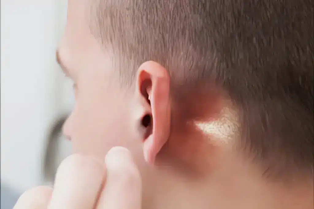
Post-Auricular Signs: The hallmark sign is a distinct, painful post-auricular swelling that actively displaces the pinna outward, downward, and forward, completely effacing the retroauricular crease. The skin overlying the mastoid process is erythematous, warm, and highly tender to deep palpation.
Fluctuance: The presence of a boggy, fluctuant mass over the mastoid bone confirms the formation of a subperiosteal abscess.
Otoscopy: Otoscopic examination reveals a bulging, intensely erythematous, and immobile tympanic membrane, or active purulent otorrhea if a perforation is present. A highly specific otoscopic sign of coalescent mastoiditis is the sagging or drooping of the postero-superior external auditory canal wall, driven by the intense inflammation of the adjacent anterior mastoid wall.
Monitoring for Complications
Clinicians must evaluate every patient for systemic and intracranial red flags. These include a high-riding, swinging fever; signs of meningeal irritation such as neck stiffness, Kernig's sign, or Brudzinski's sign; new-onset focal neurological deficits; cranial nerve palsies, particularly the abducens (sixth) nerve in Gradenigo's syndrome or the facial (seventh) nerve; papilledema; projectile vomiting; or a fluctuating level of consciousness.
History
Patients or caregivers typically report a recent or ongoing episode of acute otitis media, presenting with intense ear pain, high-grade fever, irritability, poor feeding, and lethargy. In many cases, symptoms persist or worsen despite a course of oral antibiotics prescribed for the initial ear infection.
Physical Examination
The physical findings of acute mastoiditis are highly characteristic:
Post-Auricular Signs: The hallmark sign is a distinct, painful post-auricular swelling that actively displaces the pinna outward, downward, and forward, completely effacing the retroauricular crease. The skin overlying the mastoid process is erythematous, warm, and highly tender to deep palpation.
Fluctuance: The presence of a boggy, fluctuant mass over the mastoid bone confirms the formation of a subperiosteal abscess.
Otoscopy: Otoscopic examination reveals a bulging, intensely erythematous, and immobile tympanic membrane, or active purulent otorrhea if a perforation is present. A highly specific otoscopic sign of coalescent mastoiditis is the sagging or drooping of the postero-superior external auditory canal wall, driven by the intense inflammation of the adjacent anterior mastoid wall.
Monitoring for Complications
Clinicians must evaluate every patient for systemic and intracranial red flags. These include a high-riding, swinging fever; signs of meningeal irritation such as neck stiffness, Kernig's sign, or Brudzinski's sign; new-onset focal neurological deficits; cranial nerve palsies, particularly the abducens (sixth) nerve in Gradenigo's syndrome or the facial (seventh) nerve; papilledema; projectile vomiting; or a fluctuating level of consciousness.
4
Diagnosis & Workup
2 min•227 words
The diagnosis of acute mastoiditis is primarily established via clinical examination, but a protocolized diagnostic workup must be initiated immediately to confirm the extent of the disease and plan surgical interventions.
Laboratory Workup: Request an immediate complete blood count, which typically reveals a marked leukocytosis with a left shift. Check acute-phase reactants, including C-Reactive Protein and the Erythrocyte Sedimentation Rate, which are heavily elevated.
Microbiological Workup: If the tympanic membrane is actively perforating or a drainage procedure is performed, obtain a high-quality sample of the purulent otorrhea immediately for Gram staining, culture, and targeted antibiotic sensitivity testing. Blood cultures should be drawn for all patients presenting with high-grade, spiking fevers or signs of systemic sepsis.
Contrast-Enhanced Computed Tomography (CT) Scan: A high-resolution contrast-enhanced CT scan of the temporal bones and brain is the gold standard diagnostic modality. A CT scan must be ordered immediately if there is any clinical ambiguity, suspicion of coalescent mastoiditis, evidence of a subperiosteal abscess, or any clinical red flags indicating intracranial extension.
The classic CT features of acute mastoiditis include complete opacification of the middle ear and mastoid air cells due to fluid and mucosal swelling, accompanied by the clear loss or dissolution of the fine, bony intracellular septa. Contrast-enhanced scans can simultaneously rule out concurrent intracranial complications, such as an epidural abscess, subdural empyema, meningitis, brain abscess, or sigmoid sinus thrombosis.
Laboratory Workup: Request an immediate complete blood count, which typically reveals a marked leukocytosis with a left shift. Check acute-phase reactants, including C-Reactive Protein and the Erythrocyte Sedimentation Rate, which are heavily elevated.
Microbiological Workup: If the tympanic membrane is actively perforating or a drainage procedure is performed, obtain a high-quality sample of the purulent otorrhea immediately for Gram staining, culture, and targeted antibiotic sensitivity testing. Blood cultures should be drawn for all patients presenting with high-grade, spiking fevers or signs of systemic sepsis.
Contrast-Enhanced Computed Tomography (CT) Scan: A high-resolution contrast-enhanced CT scan of the temporal bones and brain is the gold standard diagnostic modality. A CT scan must be ordered immediately if there is any clinical ambiguity, suspicion of coalescent mastoiditis, evidence of a subperiosteal abscess, or any clinical red flags indicating intracranial extension.
The classic CT features of acute mastoiditis include complete opacification of the middle ear and mastoid air cells due to fluid and mucosal swelling, accompanied by the clear loss or dissolution of the fine, bony intracellular septa. Contrast-enhanced scans can simultaneously rule out concurrent intracranial complications, such as an epidural abscess, subdural empyema, meningitis, brain abscess, or sigmoid sinus thrombosis.
5
Management
3 min•472 words
The management of acute mastoiditis must be aggressive, inpatient-based, and highly collaborative, combining targeted intravenous pharmacotherapy with timely surgical drainage.
1. Intravenous Antibiotic Therapy
Empiric high-dose intravenous antibiotic therapy must be initiated immediately upon admission. The chosen regimen must provide broad-spectrum cover against Streptococcus pneumoniae and other common otopathogens, and must effectively cross the blood-brain barrier.
A standard first-line empiric regimen consists of a third-generation cephalosporin, such as Intravenous Ceftriaxone (80 mg/kg/day in children divided 12-to-24 hourly, or 2g 12-hourly in adults). If a chronic or pseudomonal infection is suspected, or if the patient has a history of recurrent otitis media, an anti-pseudomonal beta-lactam such as Ceftazidime or Piperacillin-Tazobactam should be selected instead.
To cover resistant Gram-positive organisms like Methicillin-Resistant Staphylococcus aureus (MRSA), Intravenous Vancomycin can be added to the primary regimen. This empiric therapy must be refined as soon as definitive culture and sensitivity results become available, and intravenous antibiotics must be maintained for at least 7 to 14 days, followed by a tailored course of oral antibiotics for a total treatment duration of 3 to 4 weeks.
2. Surgical and Drainage Interventions
Medical therapy alone is frequently insufficient for advanced or coalescent stages of mastoiditis due to the poor penetration of antibiotics into a walled-off empyema.
Myringotomy and Tympanostomy Tube Insertion: All patients presenting with acute mastoiditis who have an intact, bulging tympanic membrane should undergo an urgent myringotomy. Making a targeted incision in the tympanic membrane allows immediate decompression of the middle ear cleft, relieves excruciating pain, provides high-yield purulent material for culture, and restores ventilation. The placement of a tympanostomy tube (grommet) ensures continuous, long-term middle ear drainage.
Incision and Drainage: If a fluctuant post-auricular subperiosteal abscess is present, urgent surgical incision and drainage must be performed to evacuate the purulent collection and prevent overlying skin necrosis.
Cortical Mastoidectomy: A cortical mastoidectomy involves the formal surgical opening of the mastoid cortex and the systematic exenteration of all infected, fluid-filled, and necrotic bony intercellular septa, transforming the mastoid process into a clean, open cavity while preserving the integrity of the middle ear ossicles.
A cortical mastoidectomy is definitively indicated if the patient fails to show significant clinical improvement (resolution of fever, decreased pain, and lessening swelling) within 24 to 48 hours of starting appropriate intravenous antibiotics; if there is clear CT evidence of coalescent mastoiditis or cortical bone erosion; or if the patient presents with or develops any signs of intracranial complications.
3. Supportive and Neuro-Intensive Care
Provide aggressive supportive care, including protocolized intravenous fluid hydration to correct deficits from poor oral intake, and scheduled analgesics (such as Paracetamol and Ibuprofen) to control severe otalgia.
For patients displaying signs of intracranial extension, continuous, high-dependency neurological monitoring is mandatory, tracking pupillary reflexes, Glasgow Coma Scale scores, and focal deficits hourly to identify signs of shifting intracranial pressure or impending herniation.
1. Intravenous Antibiotic Therapy
Empiric high-dose intravenous antibiotic therapy must be initiated immediately upon admission. The chosen regimen must provide broad-spectrum cover against Streptococcus pneumoniae and other common otopathogens, and must effectively cross the blood-brain barrier.
A standard first-line empiric regimen consists of a third-generation cephalosporin, such as Intravenous Ceftriaxone (80 mg/kg/day in children divided 12-to-24 hourly, or 2g 12-hourly in adults). If a chronic or pseudomonal infection is suspected, or if the patient has a history of recurrent otitis media, an anti-pseudomonal beta-lactam such as Ceftazidime or Piperacillin-Tazobactam should be selected instead.
To cover resistant Gram-positive organisms like Methicillin-Resistant Staphylococcus aureus (MRSA), Intravenous Vancomycin can be added to the primary regimen. This empiric therapy must be refined as soon as definitive culture and sensitivity results become available, and intravenous antibiotics must be maintained for at least 7 to 14 days, followed by a tailored course of oral antibiotics for a total treatment duration of 3 to 4 weeks.
2. Surgical and Drainage Interventions
Medical therapy alone is frequently insufficient for advanced or coalescent stages of mastoiditis due to the poor penetration of antibiotics into a walled-off empyema.
Myringotomy and Tympanostomy Tube Insertion: All patients presenting with acute mastoiditis who have an intact, bulging tympanic membrane should undergo an urgent myringotomy. Making a targeted incision in the tympanic membrane allows immediate decompression of the middle ear cleft, relieves excruciating pain, provides high-yield purulent material for culture, and restores ventilation. The placement of a tympanostomy tube (grommet) ensures continuous, long-term middle ear drainage.
Incision and Drainage: If a fluctuant post-auricular subperiosteal abscess is present, urgent surgical incision and drainage must be performed to evacuate the purulent collection and prevent overlying skin necrosis.
Cortical Mastoidectomy: A cortical mastoidectomy involves the formal surgical opening of the mastoid cortex and the systematic exenteration of all infected, fluid-filled, and necrotic bony intercellular septa, transforming the mastoid process into a clean, open cavity while preserving the integrity of the middle ear ossicles.
A cortical mastoidectomy is definitively indicated if the patient fails to show significant clinical improvement (resolution of fever, decreased pain, and lessening swelling) within 24 to 48 hours of starting appropriate intravenous antibiotics; if there is clear CT evidence of coalescent mastoiditis or cortical bone erosion; or if the patient presents with or develops any signs of intracranial complications.
3. Supportive and Neuro-Intensive Care
Provide aggressive supportive care, including protocolized intravenous fluid hydration to correct deficits from poor oral intake, and scheduled analgesics (such as Paracetamol and Ibuprofen) to control severe otalgia.
For patients displaying signs of intracranial extension, continuous, high-dependency neurological monitoring is mandatory, tracking pupillary reflexes, Glasgow Coma Scale scores, and focal deficits hourly to identify signs of shifting intracranial pressure or impending herniation.
6
Key Pearls & Takeaways
1 min•196 words
Coalescence Demands Clearance: Acute mastoiditis is not a simple soft-tissue infection. Once the internal bony septa dissolve (coalescent mastoiditis), an empyema is established that cannot be cleared by antibiotics alone; it requires surgical decompression and drainage.
Beware the Sagging Canal: Pay close attention to the external auditory canal during otoscopy. Sagging or drooping of the postero-superior canal wall is a pathognomonic physical sign of advanced mastoid tissue destruction.
Ear Antibiotics Mask Symptoms: A patient who develops acute mastoiditis while already taking oral antibiotics for acute otitis media may present without a high fever or an obviously bulging eardrum. Rely on post-auricular tenderness, pinpoint CT imaging, and a high index of suspicion.
Never Delay the CT for Red Flags: The development of neck stiffness, a sixth or seventh cranial nerve palsy, or a fluctuating level of consciousness in a patient with a middle ear infection indicates a life-threatening intracranial extension. Order an immediate contrast-enhanced head and temporal bone CT scan.
Complete the Marathon Course: Eradicating a deep-seated bone infection requires prolonged therapy. Do not stop treatment early; maintain highly targeted antibiotic therapy for a full 3 to 4 weeks to prevent catastrophic disease recurrence or chronic osteomyelitis.
Beware the Sagging Canal: Pay close attention to the external auditory canal during otoscopy. Sagging or drooping of the postero-superior canal wall is a pathognomonic physical sign of advanced mastoid tissue destruction.
Ear Antibiotics Mask Symptoms: A patient who develops acute mastoiditis while already taking oral antibiotics for acute otitis media may present without a high fever or an obviously bulging eardrum. Rely on post-auricular tenderness, pinpoint CT imaging, and a high index of suspicion.
Never Delay the CT for Red Flags: The development of neck stiffness, a sixth or seventh cranial nerve palsy, or a fluctuating level of consciousness in a patient with a middle ear infection indicates a life-threatening intracranial extension. Order an immediate contrast-enhanced head and temporal bone CT scan.
Complete the Marathon Course: Eradicating a deep-seated bone infection requires prolonged therapy. Do not stop treatment early; maintain highly targeted antibiotic therapy for a full 3 to 4 weeks to prevent catastrophic disease recurrence or chronic osteomyelitis.
0/6