
1
Introduction
1 min•145 words
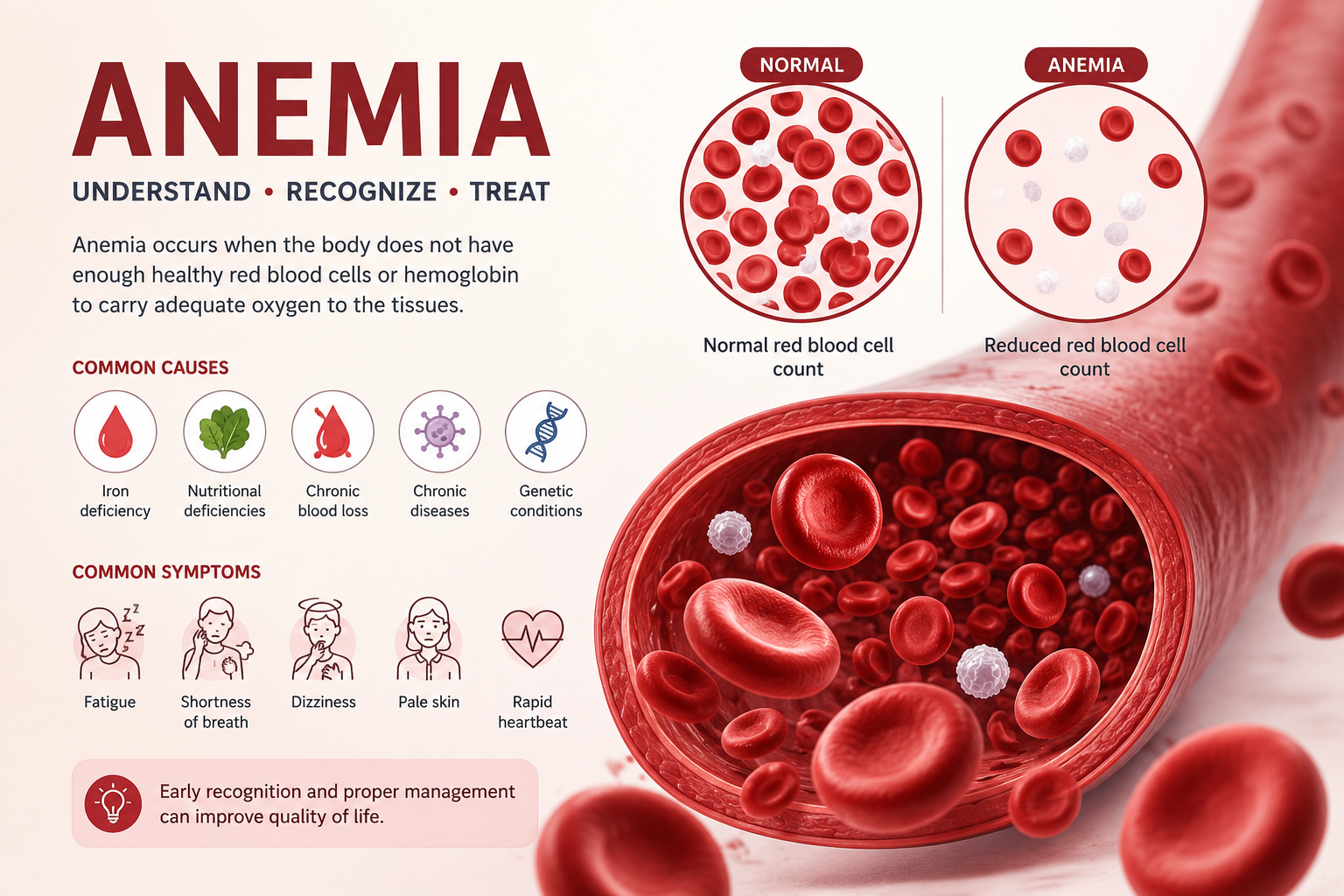
Anemia is one of the most pervasive global public health challenges, affecting more than two billion people worldwide, which constitutes roughly one-third of the global population. It is not a single disease entity but rather a critical clinical sign indicating an underlying pathological process.
Clinically, anemia is defined as a reduction in the concentration of hemoglobin, red blood cell count, or packed cell volume below established normal limits appropriate for an individual's age, gender, and physiological state, such as pregnancy. The primary functional consequence of anemia is a significant decrease in the oxygen-carrying capacity of the blood. This reduction impairs oxygen delivery to peripheral tissues, initiating a cascade of cellular hypoxia and activating various multi-system compensatory mechanisms.
In developing regions, including sub-Saharan Africa, anemia represents a massive burden of morbidity and mortality, particularly among young children, women of reproductive age, and patients with chronic co-morbidities.
Clinically, anemia is defined as a reduction in the concentration of hemoglobin, red blood cell count, or packed cell volume below established normal limits appropriate for an individual's age, gender, and physiological state, such as pregnancy. The primary functional consequence of anemia is a significant decrease in the oxygen-carrying capacity of the blood. This reduction impairs oxygen delivery to peripheral tissues, initiating a cascade of cellular hypoxia and activating various multi-system compensatory mechanisms.
In developing regions, including sub-Saharan Africa, anemia represents a massive burden of morbidity and mortality, particularly among young children, women of reproductive age, and patients with chronic co-morbidities.
2
Background & Pathophysiology
2 min•310 words
To understand the pathophysiology of anemia, clinicians must categorize it using two primary, interconnected systems: morphological classification and etiological classification.
Morphological classification is based strictly on the physical characteristics of the erythrocytes, specifically their size, measured by Mean Corpuscular Volume, and their hemoglobin concentration, measured by Mean Corpuscular Hemoglobin Concentration. This system divides anemia into three operational categories:
Microcytic Hypochromic Anemia, where the Mean Corpuscular Volume is less than 80 fL. This indicates a defect in hemoglobin synthesis, most commonly caused by iron deficiency, chronic inflammation, or globin chain synthesis disorders like Thalassemia.
Normocytic Normochromic Anemia, where the Mean Corpuscular Volume is maintained between 80 and 100 fL. This reflects conditions where red blood cell production is impaired or destruction is accelerated, but the surviving cells remain structurally normal, as seen in acute blood loss, chronic kidney disease, or early stages of chronic disease.
Macrocytic Anemia, where the Mean Corpuscular Volume exceeds 100 fL. This is further divided into megaloblastic variants, caused by impaired DNA synthesis due to Vitamin B12 or Folate deficiency, and non-megaloblastic variants, associated with alcohol use, liver disease, or hypothyroidism.
Etiological classification focuses directly on the underlying mechanism of the cell deficit:
Decreased or Impaired Red Blood Cell Production: This occurs when the bone marrow lacks raw materials due to nutritional deficiencies, lacks hormonal stimulation such as Erythropoietin deficiency in chronic kidney disease, or suffers from direct marrow suppression or aplasia.
Increased Red Blood Cell Destruction: Also known as hemolytic anemias, this group is split into intracorpuscular defects, which are structural and inherited such as Sickle Cell Disease or G6PD deficiency, and extracorpuscular defects, which are acquired via external forces like autoimmune destruction, mechanical trauma, or severe infections such as Malaria.
Absolute Blood Loss: This includes acute blood loss from trauma or surgical hemorrhage, and chronic insidious blood loss from gastrointestinal lesions, hookworm infestations, or menorrhagia.
Morphological classification is based strictly on the physical characteristics of the erythrocytes, specifically their size, measured by Mean Corpuscular Volume, and their hemoglobin concentration, measured by Mean Corpuscular Hemoglobin Concentration. This system divides anemia into three operational categories:
Microcytic Hypochromic Anemia, where the Mean Corpuscular Volume is less than 80 fL. This indicates a defect in hemoglobin synthesis, most commonly caused by iron deficiency, chronic inflammation, or globin chain synthesis disorders like Thalassemia.
Normocytic Normochromic Anemia, where the Mean Corpuscular Volume is maintained between 80 and 100 fL. This reflects conditions where red blood cell production is impaired or destruction is accelerated, but the surviving cells remain structurally normal, as seen in acute blood loss, chronic kidney disease, or early stages of chronic disease.
Macrocytic Anemia, where the Mean Corpuscular Volume exceeds 100 fL. This is further divided into megaloblastic variants, caused by impaired DNA synthesis due to Vitamin B12 or Folate deficiency, and non-megaloblastic variants, associated with alcohol use, liver disease, or hypothyroidism.
Etiological classification focuses directly on the underlying mechanism of the cell deficit:
Decreased or Impaired Red Blood Cell Production: This occurs when the bone marrow lacks raw materials due to nutritional deficiencies, lacks hormonal stimulation such as Erythropoietin deficiency in chronic kidney disease, or suffers from direct marrow suppression or aplasia.
Increased Red Blood Cell Destruction: Also known as hemolytic anemias, this group is split into intracorpuscular defects, which are structural and inherited such as Sickle Cell Disease or G6PD deficiency, and extracorpuscular defects, which are acquired via external forces like autoimmune destruction, mechanical trauma, or severe infections such as Malaria.
Absolute Blood Loss: This includes acute blood loss from trauma or surgical hemorrhage, and chronic insidious blood loss from gastrointestinal lesions, hookworm infestations, or menorrhagia.
3
Clinical Features
2 min•299 words
The clinical manifestations of anemia are driven by two main factors: the severity and speed of onset of tissue hypoxia, and the activation of the body's cardiovascular and neuroendocrine compensatory mechanisms.
Symptoms of tissue hypoxia include generalized weakness, progressive malaise, easy fatigability, and poor exercise tolerance. As oxygen delivery to the brain falls, patients report fronto-occipital headaches, vertigo, tinnitus, irritability, and an inability to concentrate. On physical examination, the hallmark sign of anemia is pallor, which is best evaluated in areas with rich superficial capillary beds, specifically the palpebral conjunctiva, the mucous membranes of the tongue and oral cavity, and the nail beds.
When anemia develops rapidly or becomes severe, the cardiovascular system attempts to compensate for low oxygen carrying capacity by inducing a hyperdynamic circulatory state. The body increases stroke volume and heart rate, leading to clinical complaints of palpitations and visible tachycardia.
On cardiac auscultation, a functional systolic ejection murmur may be heard, typically loudest at the pulmonic area, caused by increased blood flow velocity and reduced blood viscosity. If the hemoglobin falls below critical levels for a prolonged period, these compensatory systems fail, and the patient can progress into high-output congestive cardiac failure, characterized by orthopnea, elevated jugular venous pressure, hepatomegaly, and bilateral peripheral edema.
Specific etiologies also present with unique signs. For example, severe iron deficiency anemia can present with koilonychia, which are brittle, spoon-shaped nails, atrophic glossitis, which is a smooth, painful, shiny tongue, angular cheilitis, which are painful cracks at the corners of the mouth, and pica, an abnormal craving to consume non-food substances like soil, ice, or charcoal. Megaloblastic anemia due to Vitamin B12 deficiency may uniquely display neurological features, such as subacute combined degeneration of the spinal cord, manifesting as symmetrical paresthesia in a glove-and-stocking pattern, loss of proprioception, and ataxia.
Symptoms of tissue hypoxia include generalized weakness, progressive malaise, easy fatigability, and poor exercise tolerance. As oxygen delivery to the brain falls, patients report fronto-occipital headaches, vertigo, tinnitus, irritability, and an inability to concentrate. On physical examination, the hallmark sign of anemia is pallor, which is best evaluated in areas with rich superficial capillary beds, specifically the palpebral conjunctiva, the mucous membranes of the tongue and oral cavity, and the nail beds.
When anemia develops rapidly or becomes severe, the cardiovascular system attempts to compensate for low oxygen carrying capacity by inducing a hyperdynamic circulatory state. The body increases stroke volume and heart rate, leading to clinical complaints of palpitations and visible tachycardia.
On cardiac auscultation, a functional systolic ejection murmur may be heard, typically loudest at the pulmonic area, caused by increased blood flow velocity and reduced blood viscosity. If the hemoglobin falls below critical levels for a prolonged period, these compensatory systems fail, and the patient can progress into high-output congestive cardiac failure, characterized by orthopnea, elevated jugular venous pressure, hepatomegaly, and bilateral peripheral edema.
Specific etiologies also present with unique signs. For example, severe iron deficiency anemia can present with koilonychia, which are brittle, spoon-shaped nails, atrophic glossitis, which is a smooth, painful, shiny tongue, angular cheilitis, which are painful cracks at the corners of the mouth, and pica, an abnormal craving to consume non-food substances like soil, ice, or charcoal. Megaloblastic anemia due to Vitamin B12 deficiency may uniquely display neurological features, such as subacute combined degeneration of the spinal cord, manifesting as symmetrical paresthesia in a glove-and-stocking pattern, loss of proprioception, and ataxia.
4
Diagnosis & Workup
2 min•321 words
A logical and thorough laboratory workup is required to confirm anemia, evaluate its severity, and pinpoint the exact underlying etiology before starting treatment.
Complete Blood Count and Red Cell Indices: The initial step is checking the Hemoglobin concentration to classify the severity according to World Health Organization and regional guidelines. For adult men, anemia is present when hemoglobin is below 13 g/dL. For non-pregnant adult women, the threshold is below 12 g/dL, and for pregnant women, it is below 11 g/dL. Severe anemia across all adult populations is defined as a hemoglobin level below 7 to 8 g/dL depending on specific clinical guidelines. The complete blood count also provides the Mean Corpuscular Volume to guide morphological grouping, and the Red Cell Distribution Width, which measures variation in cell size, helping differentiate iron deficiency from thalassemia traits.
Peripheral Blood Film Examination: This provides a direct visual evaluation of erythrocyte morphology. It allows pathologists to confirm hypochromia, identify abnormal cell shapes such as sickle cells, target cells, or schistocytes, and look for hypersegmented neutrophils, which are a definitive sign of megaloblastic anemia.
Reticulocyte Count: The reticulocyte count is a critical marker that evaluates the functional response of the bone marrow. A low reticulocyte count indicates an underproductive marrow due to nutritional deficiencies or aplasia. A high reticulocyte count reveals an appropriately active, hyperproliferative marrow responding to ongoing peripheral destruction or recent acute blood loss.
Targeted Biochemical Workup: If microcytic anemia is identified, a full iron profile should be ordered, including serum iron, Total Iron Binding Capacity, and serum ferritin, which is the most reliable marker of depleted iron stores. If macrocytic anemia is present, serum Vitamin B12 and folate levels must be quantified. For cases showing hemolysis, clinicians must check serum lactate dehydrogenase levels, unconjugated bilirubin, haptoglobin, and perform a Direct Antiglobulin Test to exclude immune-mediated destruction. In endemic regions, a concurrent malaria rapid diagnostic test or blood smear must always be performed.
Complete Blood Count and Red Cell Indices: The initial step is checking the Hemoglobin concentration to classify the severity according to World Health Organization and regional guidelines. For adult men, anemia is present when hemoglobin is below 13 g/dL. For non-pregnant adult women, the threshold is below 12 g/dL, and for pregnant women, it is below 11 g/dL. Severe anemia across all adult populations is defined as a hemoglobin level below 7 to 8 g/dL depending on specific clinical guidelines. The complete blood count also provides the Mean Corpuscular Volume to guide morphological grouping, and the Red Cell Distribution Width, which measures variation in cell size, helping differentiate iron deficiency from thalassemia traits.
Peripheral Blood Film Examination: This provides a direct visual evaluation of erythrocyte morphology. It allows pathologists to confirm hypochromia, identify abnormal cell shapes such as sickle cells, target cells, or schistocytes, and look for hypersegmented neutrophils, which are a definitive sign of megaloblastic anemia.
Reticulocyte Count: The reticulocyte count is a critical marker that evaluates the functional response of the bone marrow. A low reticulocyte count indicates an underproductive marrow due to nutritional deficiencies or aplasia. A high reticulocyte count reveals an appropriately active, hyperproliferative marrow responding to ongoing peripheral destruction or recent acute blood loss.
Targeted Biochemical Workup: If microcytic anemia is identified, a full iron profile should be ordered, including serum iron, Total Iron Binding Capacity, and serum ferritin, which is the most reliable marker of depleted iron stores. If macrocytic anemia is present, serum Vitamin B12 and folate levels must be quantified. For cases showing hemolysis, clinicians must check serum lactate dehydrogenase levels, unconjugated bilirubin, haptoglobin, and perform a Direct Antiglobulin Test to exclude immune-mediated destruction. In endemic regions, a concurrent malaria rapid diagnostic test or blood smear must always be performed.
5
Management
2 min•372 words
The management of anemia must never be purely symptomatic. It must focus entirely on correcting the underlying cause while carefully stabilizing the patient's hemodynamic status.
Nutritional Deficiency Replacement Therapy:
For Iron Deficiency Anemia, oral iron supplementation is the primary therapy. Administer Ferrous Sulphate at a dose of 200 mg two to three times daily in adults, or elemental iron calculated at 3 to 6 mg/kg/day in pediatric patients. Treatment must be continued for three to six months after the hemoglobin normalizes to completely replenish internal ferritin stores. Advise patients to take iron on an empty stomach or with Vitamin C to enhance absorption, and warn them about benign side effects like dark stools and constipation. Intravenous iron is reserved for severe malabsorption or oral intolerance.
For Vitamin B12 deficiency, administer intramuscular Hydroxycobalamin at 1000 micrograms daily or every alternate day for the first one to two weeks, followed by monthly maintenance doses. For Folate deficiency, prescribe oral Folic Acid 5 mg daily for up to four months, ensuring Vitamin B12 deficiency has been explicitly ruled out first to prevent precipitating irreversible neurological damage.
Management of Chronic Disease and Renal Anemia: When anemia is driven by chronic kidney disease, the primary issue is a lack of endogenous erythropoietin. Treatment involves the subcutaneous administration of Recombinant Human Erythropoietin or Darbepoetin alfa, tailored alongside regular intravenous iron supplements to support active red blood cell production.
Disease-Modifying Therapy in Hemolytic States: In conditions like Sickle Cell Disease, long-term management includes daily Folic Acid supplementation to support high bone marrow turnover, and Hydroxyurea therapy to stimulate the production of Fetal Hemoglobin, which prevents red cell sickling and reduces painful crises.
Judicial Blood Transfusion Protocol: Blood transfusion is an immediate, life-saving intervention but must be used judiciously. In stable, asymptomatic patients without cardiovascular disease, transfusion is generally indicated only when the hemoglobin drops below 7 g/dL. However, if the patient shows signs of symptomatic tissue hypoxia, altered mental status, active ischemic chest pain, or high-output heart failure, emergency transfusion of packed red blood cells must be initiated immediately regardless of the exact hemoglobin number. Transfusions should be given slowly, and concurrent loop diuretics like Furosemide should be considered in elderly or fluid-compromised patients to prevent transfusion-associated circulatory overload.
Nutritional Deficiency Replacement Therapy:
For Iron Deficiency Anemia, oral iron supplementation is the primary therapy. Administer Ferrous Sulphate at a dose of 200 mg two to three times daily in adults, or elemental iron calculated at 3 to 6 mg/kg/day in pediatric patients. Treatment must be continued for three to six months after the hemoglobin normalizes to completely replenish internal ferritin stores. Advise patients to take iron on an empty stomach or with Vitamin C to enhance absorption, and warn them about benign side effects like dark stools and constipation. Intravenous iron is reserved for severe malabsorption or oral intolerance.
For Vitamin B12 deficiency, administer intramuscular Hydroxycobalamin at 1000 micrograms daily or every alternate day for the first one to two weeks, followed by monthly maintenance doses. For Folate deficiency, prescribe oral Folic Acid 5 mg daily for up to four months, ensuring Vitamin B12 deficiency has been explicitly ruled out first to prevent precipitating irreversible neurological damage.
Management of Chronic Disease and Renal Anemia: When anemia is driven by chronic kidney disease, the primary issue is a lack of endogenous erythropoietin. Treatment involves the subcutaneous administration of Recombinant Human Erythropoietin or Darbepoetin alfa, tailored alongside regular intravenous iron supplements to support active red blood cell production.
Disease-Modifying Therapy in Hemolytic States: In conditions like Sickle Cell Disease, long-term management includes daily Folic Acid supplementation to support high bone marrow turnover, and Hydroxyurea therapy to stimulate the production of Fetal Hemoglobin, which prevents red cell sickling and reduces painful crises.
Judicial Blood Transfusion Protocol: Blood transfusion is an immediate, life-saving intervention but must be used judiciously. In stable, asymptomatic patients without cardiovascular disease, transfusion is generally indicated only when the hemoglobin drops below 7 g/dL. However, if the patient shows signs of symptomatic tissue hypoxia, altered mental status, active ischemic chest pain, or high-output heart failure, emergency transfusion of packed red blood cells must be initiated immediately regardless of the exact hemoglobin number. Transfusions should be given slowly, and concurrent loop diuretics like Furosemide should be considered in elderly or fluid-compromised patients to prevent transfusion-associated circulatory overload.
6
Key Pearls & Takeaways
1 min•194 words
Anemia is a Sign, Not a Diagnosis: Never simply write a prescription for iron or order a transfusion without identifying the primary cause. Anemia is always a manifestation of an underlying pathology that requires clear diagnosis.
Ferritin is the Gold Standard: When evaluating suspected iron deficiency, a low serum ferritin level is the most specific and accurate biomarker of depleted body iron stores, even before red cell indices drop.
The B12 and Folate Trap: Never treat a macrocytic anemia with high-dose folic acid alone without checking Vitamin B12 levels. Folic acid will correct the hematological parameters but will allow the neurological damage of B12 deficiency to progress unchecked.
Transfuse the Patient, Not the Lab Number: A hemoglobin level of 6 g/dL in a young, walking, asymptomatic patient may be managed with oral medications, while a hemoglobin level of 8 g/dL in an elderly patient with crushing chest pain and heart failure requires an immediate blood transfusion.
Complete the Course: Oral iron therapy must not be stopped as soon as the hemoglobin reaches normal limits. It must be continued strictly for an additional three to six months to completely refill the body's structural iron stores.
Ferritin is the Gold Standard: When evaluating suspected iron deficiency, a low serum ferritin level is the most specific and accurate biomarker of depleted body iron stores, even before red cell indices drop.
The B12 and Folate Trap: Never treat a macrocytic anemia with high-dose folic acid alone without checking Vitamin B12 levels. Folic acid will correct the hematological parameters but will allow the neurological damage of B12 deficiency to progress unchecked.
Transfuse the Patient, Not the Lab Number: A hemoglobin level of 6 g/dL in a young, walking, asymptomatic patient may be managed with oral medications, while a hemoglobin level of 8 g/dL in an elderly patient with crushing chest pain and heart failure requires an immediate blood transfusion.
Complete the Course: Oral iron therapy must not be stopped as soon as the hemoglobin reaches normal limits. It must be continued strictly for an additional three to six months to completely refill the body's structural iron stores.
0/6