
1
Introduction
1 min•140 words
Viral Hemorrhagic Fevers (VHFs) encompass a group of severe, life-threatening zoonotic infections caused by distinct families of RNA viruses. These diseases are characterized by a sudden onset of a febrile syndrome, progressive multi-organ dysfunction, variable bleeding manifestations, and hypovolemic shock. VHFs are notorious for causing major epidemics associated with exceptionally high case fatality rates.
Historically, these outbreaks have been severely exacerbated by a lack of specific medical countermeasures, a shortage of timely laboratory diagnostics, late clinical detection, and inadequate infection control practices within local healthcare facilities. Among these pathogens, Bundibugyo Virus Disease (BDBV) stands as a critical species within the Ebolavirus genus. First discovered in 2007 during a severe outbreak in the Bundibugyo district of Western Uganda, BDBV highlights the persistent threat of spillover epidemics in equatorial Africa and demands a high index of clinical suspicion and rigorous institutional containment.
Historically, these outbreaks have been severely exacerbated by a lack of specific medical countermeasures, a shortage of timely laboratory diagnostics, late clinical detection, and inadequate infection control practices within local healthcare facilities. Among these pathogens, Bundibugyo Virus Disease (BDBV) stands as a critical species within the Ebolavirus genus. First discovered in 2007 during a severe outbreak in the Bundibugyo district of Western Uganda, BDBV highlights the persistent threat of spillover epidemics in equatorial Africa and demands a high index of clinical suspicion and rigorous institutional containment.
2
Background & Pathophysiology
2 min•378 words
To understand the operational dynamics of VHFs, clinicians must comprehend both the taxonomy of the causative viral families and the ecological lifecycles that drive human spillover.
Taxonomy of VHF Viral Families
The primary viruses responsible for hemorrhagic fevers are grouped into four distinct taxonomic families:
Filoviridae: Includes the Ebolavirus genus (comprising Ebola Zaire [EBOV], Ebola Sudan [SUDV], and Ebola Bundibugyo [BDBV]), the Marburgvirus genus, and the Cuevavirus genus.
Arenaviridae: Responsible for Lassa Hemorrhagic Fever and various South American hemorrhagic fevers (Argentine, Bolivian, Venezuelan, and Brazilian HFs).
Bunyaviridae: Includes the Hantavirus genus, Crimean-Congo Hemorrhagic Fever (CCHF), and Rift Valley Fever (RVF).
Flaviviridae: Includes Yellow Fever, Dengue Hemorrhagic Fever, Kyasanur Forest Disease, and Omsk Hemorrhagic Fever.
Ebolavirus Ecology and Structural Fragility
The structural biology of the Ebolavirus reveals a prominent outer lipid envelope. Because of this lipid membrane, the virus is highly fragile outside the host organism and is easily destroyed by standard soap, household chlorine solutions, and direct environmental exposure.
In nature, the virus oscillates between two distinct cycles:
Enzootic Cycle: Evidence strongly implicates fruit bats of the Pteropodidae family as the primary reservoir hosts. The virus is maintained within these bat populations through mechanisms that are not yet fully understood.
Epizootic Cycle: Sporadic epizootics occur among non-human primates (such as chimpanzees and gorillas) and forest duikers. These animals suffer high mortality rates from the infection, and these die-offs frequently precede human epidemics.
Amplification and Human Transmission Pathways
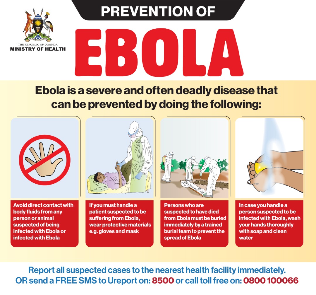
An epidemic is triggered by an initial human spillover event resulting from direct contact with the blood, secretions, or organs of an infected bat or wild animal. Once introduced into the human population, interhuman amplification occurs via:
Primary Modes: Direct contact with the blood, body fluids (saliva, vomitus, stool, semen, sweat, breast milk), or organs of an infected, symptomatic individual.
Secondary Modes: Exposure to contaminated fomites, such as needles, soiled bed linens, surfaces, or medical equipment.
Vertical Transmission: Mother-to-child transmission routes via the placenta, during delivery, or through breast milk.
This transmission is heavily amplified in two settings: within communities via family members caring for the sick and unsafe funeral or burial practices involving direct contact with the deceased; and within healthcare facilities due to inadequate infection control, unsafe injections, and exposure of healthcare workers to patients before isolation is established.
Taxonomy of VHF Viral Families
The primary viruses responsible for hemorrhagic fevers are grouped into four distinct taxonomic families:
Filoviridae: Includes the Ebolavirus genus (comprising Ebola Zaire [EBOV], Ebola Sudan [SUDV], and Ebola Bundibugyo [BDBV]), the Marburgvirus genus, and the Cuevavirus genus.
Arenaviridae: Responsible for Lassa Hemorrhagic Fever and various South American hemorrhagic fevers (Argentine, Bolivian, Venezuelan, and Brazilian HFs).
Bunyaviridae: Includes the Hantavirus genus, Crimean-Congo Hemorrhagic Fever (CCHF), and Rift Valley Fever (RVF).
Flaviviridae: Includes Yellow Fever, Dengue Hemorrhagic Fever, Kyasanur Forest Disease, and Omsk Hemorrhagic Fever.
Ebolavirus Ecology and Structural Fragility
The structural biology of the Ebolavirus reveals a prominent outer lipid envelope. Because of this lipid membrane, the virus is highly fragile outside the host organism and is easily destroyed by standard soap, household chlorine solutions, and direct environmental exposure.
In nature, the virus oscillates between two distinct cycles:
Enzootic Cycle: Evidence strongly implicates fruit bats of the Pteropodidae family as the primary reservoir hosts. The virus is maintained within these bat populations through mechanisms that are not yet fully understood.
Epizootic Cycle: Sporadic epizootics occur among non-human primates (such as chimpanzees and gorillas) and forest duikers. These animals suffer high mortality rates from the infection, and these die-offs frequently precede human epidemics.
Amplification and Human Transmission Pathways
An epidemic is triggered by an initial human spillover event resulting from direct contact with the blood, secretions, or organs of an infected bat or wild animal. Once introduced into the human population, interhuman amplification occurs via:
Primary Modes: Direct contact with the blood, body fluids (saliva, vomitus, stool, semen, sweat, breast milk), or organs of an infected, symptomatic individual.
Secondary Modes: Exposure to contaminated fomites, such as needles, soiled bed linens, surfaces, or medical equipment.
Vertical Transmission: Mother-to-child transmission routes via the placenta, during delivery, or through breast milk.
This transmission is heavily amplified in two settings: within communities via family members caring for the sick and unsafe funeral or burial practices involving direct contact with the deceased; and within healthcare facilities due to inadequate infection control, unsafe injections, and exposure of healthcare workers to patients before isolation is established.
3
Clinical Features
2 min•280 words
The incubation period for VHFs, particularly filoviruses like BDBV, ranges strictly from 2 to 21 days. It is a vital clinical rule that humans are completely non-infectious until they develop symptoms. Monitoring exposed contacts must be strictly maintained for the duration of this 21-day window.
The clinical presentation of Ebola Virus Disease (EVD) progresses through three distinct, overlapping clinical stages:
First Stage (Early Non-Specific Phase)
Patients typically present in this initial phase, where symptoms are completely non-specific and easily confused with endemic diseases like malaria or typhoid. Signs include a sudden onset of high fever, profound body weakness, severe fronto-occipital headaches, intense joint and back pain, generalized myalgia, severe sore throat, chest pain, conjunctival injection (eye redness), and a maculopapular skin rash.
Second Stage (Gastrointestinal Phase)
As the disease progresses into the second phase, the virus causes extensive damage to the gastrointestinal mucosa. The patient develops severe abdominal pain, persistent vomiting, profuse watery diarrhea, and worsening dysphagia (difficulty swallowing). Hiccups may also develop during this stage, serving as a poor prognostic indicator.
Advanced Stage (Organ Failure and Hemorrhagic Phase)
The advanced stage is characterized by multi-organ system collapse. Manifestations include altered mental status, confusion, delirium, somnolence, convulsions, seizures, and coma. Respiratory distress and rapid breathing (tachypnea) signal underlying pulmonary injury. Active hemorrhage occurs due to severe consumptive coagulopathy and endothelial dysfunction.
It is a critical clinical pearl that frank hemorrhage is a late symptom and is not present in all patients; it is seen in only approximately 40% of EVD or Marburg cases, and 15% or fewer in Rift Valley Fever or CCHF cases. The terminal phase involves profound hypovolemic and distributive shock, followed by complete multi-organ failure and death.
The clinical presentation of Ebola Virus Disease (EVD) progresses through three distinct, overlapping clinical stages:
First Stage (Early Non-Specific Phase)
Patients typically present in this initial phase, where symptoms are completely non-specific and easily confused with endemic diseases like malaria or typhoid. Signs include a sudden onset of high fever, profound body weakness, severe fronto-occipital headaches, intense joint and back pain, generalized myalgia, severe sore throat, chest pain, conjunctival injection (eye redness), and a maculopapular skin rash.
Second Stage (Gastrointestinal Phase)
As the disease progresses into the second phase, the virus causes extensive damage to the gastrointestinal mucosa. The patient develops severe abdominal pain, persistent vomiting, profuse watery diarrhea, and worsening dysphagia (difficulty swallowing). Hiccups may also develop during this stage, serving as a poor prognostic indicator.
Advanced Stage (Organ Failure and Hemorrhagic Phase)
The advanced stage is characterized by multi-organ system collapse. Manifestations include altered mental status, confusion, delirium, somnolence, convulsions, seizures, and coma. Respiratory distress and rapid breathing (tachypnea) signal underlying pulmonary injury. Active hemorrhage occurs due to severe consumptive coagulopathy and endothelial dysfunction.
It is a critical clinical pearl that frank hemorrhage is a late symptom and is not present in all patients; it is seen in only approximately 40% of EVD or Marburg cases, and 15% or fewer in Rift Valley Fever or CCHF cases. The terminal phase involves profound hypovolemic and distributive shock, followed by complete multi-organ failure and death.
4
Diagnosis & Workup
2 min•370 words
Clinicians must maintain an exceptionally high index of suspicion in an outbreak setting. Because early manifestations are indistinguishable from other tropical fevers, diagnosis requires a meticulous history and a structured epidemiological workup.
History and Epidemiological Links
History taking must aggressively explore:
The exact timeline of the patient's symptoms.
Detailed travel history to known outbreak regions.
Any direct or indirect contact with individuals displaying similar febrile illnesses, unexplained bleeding, or history of sudden death.
Any occupational exposures, such as hunting, processing bushmeat, entering bat-infested caves or mines (associated with Marburg and Ebola), or handling sick livestock (associated with Rift Valley Fever and Crimean-Congo Hemorrhagic Fever).
Clinical history noting febrile conditions that have failed to respond to standard regional treatments, such as first-line antimalarials or broad-spectrum antibiotics.
Case Definitions
To guide operational management, cases are stratified using standardized clinical metrics:
Suspect Case: Evaluated using standard criteria:
Any contact with a known case who develops a fever.
Any contact with a known case who exhibits one or more specific VHF symptoms.
Any individual presenting with a fever accompanied by three or more classic symptoms (e.g., headache, vomiting, diarrhea, myalgia).
Any individual presenting with inexplicable, spontaneous bleeding from any site.
Any inexplicable or sudden death in the community.
Confirmed Case: Any suspected case with a definitive positive laboratory diagnosis, primarily achieved via a positive Reverse Transcription Polymerase Chain Reaction (RT-PCR) test.
Differential Diagnosis and Minimal Workup
The primary differential diagnoses that must be excluded include:
Malaria (the most critical mimic; a rapid diagnostic test [mRDT] must be performed immediately for all febrile patients).
Typhoid fever, Shigellosis, and Cholera (for gastrointestinal presentations).
Dengue fever, Leptospirosis, Meningitis, Hepatitis, acute HIV, and disseminated Tuberculosis.
Laboratory Abnormalities
In specialized or isolation settings where laboratory workups can be safely performed under strict biocontainment, common abnormalities include:
Early Phase: Severe electrolyte shifts (derangements in Potassium, Sodium, Magnesium, and Bicarbonate) due to gastrointestinal fluid losses.
Late Phase (Shock/Multi-Organ Failure): Markedly elevated Serum Creatinine and Blood Urea Nitrogen (BUN) indicating Acute Kidney Injury; significantly elevated Lactic Acid confirming tissue hypoperfusion; highly elevated transaminases (AST and ALT, with AST typically exceeding ALT); altered Blood Glucose levels; and prolonged coagulation profiles, including elevated Partial Thromboplastin Time (PTT) and an increased International Normalized Ratio (INR).
History and Epidemiological Links
History taking must aggressively explore:
The exact timeline of the patient's symptoms.
Detailed travel history to known outbreak regions.
Any direct or indirect contact with individuals displaying similar febrile illnesses, unexplained bleeding, or history of sudden death.
Any occupational exposures, such as hunting, processing bushmeat, entering bat-infested caves or mines (associated with Marburg and Ebola), or handling sick livestock (associated with Rift Valley Fever and Crimean-Congo Hemorrhagic Fever).
Clinical history noting febrile conditions that have failed to respond to standard regional treatments, such as first-line antimalarials or broad-spectrum antibiotics.
Case Definitions
To guide operational management, cases are stratified using standardized clinical metrics:
Suspect Case: Evaluated using standard criteria:
Any contact with a known case who develops a fever.
Any contact with a known case who exhibits one or more specific VHF symptoms.
Any individual presenting with a fever accompanied by three or more classic symptoms (e.g., headache, vomiting, diarrhea, myalgia).
Any individual presenting with inexplicable, spontaneous bleeding from any site.
Any inexplicable or sudden death in the community.
Confirmed Case: Any suspected case with a definitive positive laboratory diagnosis, primarily achieved via a positive Reverse Transcription Polymerase Chain Reaction (RT-PCR) test.
Differential Diagnosis and Minimal Workup
The primary differential diagnoses that must be excluded include:
Malaria (the most critical mimic; a rapid diagnostic test [mRDT] must be performed immediately for all febrile patients).
Typhoid fever, Shigellosis, and Cholera (for gastrointestinal presentations).
Dengue fever, Leptospirosis, Meningitis, Hepatitis, acute HIV, and disseminated Tuberculosis.
Laboratory Abnormalities
In specialized or isolation settings where laboratory workups can be safely performed under strict biocontainment, common abnormalities include:
Early Phase: Severe electrolyte shifts (derangements in Potassium, Sodium, Magnesium, and Bicarbonate) due to gastrointestinal fluid losses.
Late Phase (Shock/Multi-Organ Failure): Markedly elevated Serum Creatinine and Blood Urea Nitrogen (BUN) indicating Acute Kidney Injury; significantly elevated Lactic Acid confirming tissue hypoperfusion; highly elevated transaminases (AST and ALT, with AST typically exceeding ALT); altered Blood Glucose levels; and prolonged coagulation profiles, including elevated Partial Thromboplastin Time (PTT) and an increased International Normalized Ratio (INR).
5
Management
5 min•875 words
The management of VHFs combines aggressive supportive care with the utilization of specific approved therapeutics where available, always executed within a strict isolation framework.
Therapeutics and Vaccine Availability Summary
The availability of specific countermeasures varies significantly by pathogen species:
Ebola Zaire (EBOV): Approved therapeutics include specific monoclonal antibodies, namely mAb114 (Ansuvimab) and REGN-EB3 (Inmazeb), which have been proven safe for use in both pregnancy and lactation. Conversely, alternative agents like ZMapp and Remdesivir are explicitly not recommended for EBOV treatment. Preventive vaccines are available, including Ervebo (a single-dose vaccine) and the Zabdeno/Mvabea two-dose regimen.
Ebola Sudan (SUDV) and Ebola Bundibugyo (BDBV): There are currently no approved specific therapeutics or vaccines. Interventions are administered strictly under Monitored Emergency Use of Unregistered and Investigational Interventions (MEURI) or compassionate use protocols, which may include investigational agents like Remdesivir or the MBP134 monoclonal antibody cocktail. Candidate vaccines are in active development.
Marburg Virus Disease (MVD): No approved specific treatments or vaccines exist. Experimental monoclonal antibodies and small-molecule antivirals are restricted to MEURI protocols while candidate vaccines undergo clinical trials.
Crimean-Congo Hemorrhagic Fever (CCHF): No specific approved treatments or vaccines are available, though the antiviral drug Ribavirin is frequently administered off-label with noted clinical benefits if started very early in the course of the illness.
Rift Valley Fever (RVF): Management is entirely supportive; no specific approved therapeutics or vaccines are available for human use.
Handling of Suspected Cases and Isolation
Any patient matching the suspect case definition must be isolated immediately within a functional isolation area. Large-volume healthcare facilities must maintain a dedicated spatial layout for this purpose.
Transfer to a specialized Isolation Unit (IDU) must be arranged promptly using dedicated transport teams. Healthcare workers who experience an accidental exposure or needle-stick injury must practice immediate self-isolation and report the event to the institutional safety officer.
Discharging Patients from the Isolation Unit
A recovered patient can only be safely discharged from the IDU when they fulfill all four of the following criteria:
Free of fever and significant clinical symptoms (vomiting, diarrhea, hemorrhage) for three or more consecutive days.
Demonstrating significant and sustained improvement in their overall clinical condition.
Fully capable of performing basic Activities of Daily Living (ADLs) independently.
Achieving at least one negative blood RT-PCR test result. If a clinically recovered patient remains PCR positive, the test must be repeated every 48 hours until clear, as viral RNA can persist in the blood for several days after symptoms resolve.
Post-Discharge Survivor Counselling Guidelines
Discharged survivors require extensive behavioral counseling due to persistent viral replication in immunologically privileged sites:
Male Survivors: Active virus can remain viable in semen for up to 18.5 months, and viral RNA has been detected beyond two years post-recovery. Men must undergo monthly semen surveillance and testing until they achieve two consecutive negative semen PCR results. They must be counseled extensively on safe sex practices and the mandatory use of barrier methods (condoms) until clearance is documented.
Female Survivors: Women face an extremely high risk of miscarriage, spontaneous abortion, and fetal death. A pregnant survivor who subsequently miscarries is automatically classified as an active suspect VHF case and must be readmitted to the IDU until her products of conception and blood test negative by PCR. If breast milk tests positive for the virus, breastfeeding must be suspended, and the milk must be re-tested every 48 hours until a negative result is obtained.
Infection Prevention & Control (IPC)
Controlling a VHF outbreak requires strict adherence to the Chain of Infection within healthcare facilities. This involves breaking the links between the Infectious Agent, the Reservoir, the Portal of Exit, the Means of Transmission, the Portal of Entry, and the Susceptible Host. This is operationalized through two core frameworks: the IPC Hierarchy of Controls and Standard Precautions.
The IPC Hierarchy of Controls
The structural layers of defense are organized as an inverted pyramid:
Elimination: The most effective layer; physically removes the hazard through rigorous patient screening at facility entry points, reducing entry pathways, and strictly restricting visitors.
Substitution: Finding alternative clinical workflows or care methods that minimize the baseline potential for viral transmission.
Engineering Controls: Implementing physical infrastructure barriers, negative-pressure isolation zones, and dedicated spatial layouts to completely segregate clean areas from contaminated zones and restrict staff and patient movement.
Administrative Controls: Changing how personnel work by enforcing strict Standard Operating Procedures (SOPs), implementing mandatory specialized staff training, and modifying staffing ratios to prevent provider fatigue.
Personal Protective Equipment (PPE): The final, most vulnerable line of personal defense. PPE must be worn strictly according to a rapid risk assessment, ensuring full body coverage (fluid-resistant gowns, double gloving, face shields, respirators or masks, and waterproof boots) whenever contact with an isolation area is required.
Standard and Transmission-Based Precautions
Standard Precautions must be universally applied to all patients at all times, regardless of their suspected status. These include strict hand hygiene (using alcohol-based hand rubs or clean water and soap), mandatory injection safety, sharps injury prevention protocols, safe segregation and disposal of healthcare waste, proper handling of soiled linens, thorough environmental cleaning using appropriate chlorine dilutions, sterilization of patient-care equipment, and proper respiratory hygiene.
Transmission-Based Precautions combine Standard Precautions with explicit Contact and Droplet precautions to completely halt the transmission of highly infectious bodily fluids and droplets within the clinical environment.
Therapeutics and Vaccine Availability Summary
The availability of specific countermeasures varies significantly by pathogen species:
Ebola Zaire (EBOV): Approved therapeutics include specific monoclonal antibodies, namely mAb114 (Ansuvimab) and REGN-EB3 (Inmazeb), which have been proven safe for use in both pregnancy and lactation. Conversely, alternative agents like ZMapp and Remdesivir are explicitly not recommended for EBOV treatment. Preventive vaccines are available, including Ervebo (a single-dose vaccine) and the Zabdeno/Mvabea two-dose regimen.
Ebola Sudan (SUDV) and Ebola Bundibugyo (BDBV): There are currently no approved specific therapeutics or vaccines. Interventions are administered strictly under Monitored Emergency Use of Unregistered and Investigational Interventions (MEURI) or compassionate use protocols, which may include investigational agents like Remdesivir or the MBP134 monoclonal antibody cocktail. Candidate vaccines are in active development.
Marburg Virus Disease (MVD): No approved specific treatments or vaccines exist. Experimental monoclonal antibodies and small-molecule antivirals are restricted to MEURI protocols while candidate vaccines undergo clinical trials.
Crimean-Congo Hemorrhagic Fever (CCHF): No specific approved treatments or vaccines are available, though the antiviral drug Ribavirin is frequently administered off-label with noted clinical benefits if started very early in the course of the illness.
Rift Valley Fever (RVF): Management is entirely supportive; no specific approved therapeutics or vaccines are available for human use.
Handling of Suspected Cases and Isolation
Any patient matching the suspect case definition must be isolated immediately within a functional isolation area. Large-volume healthcare facilities must maintain a dedicated spatial layout for this purpose.
Transfer to a specialized Isolation Unit (IDU) must be arranged promptly using dedicated transport teams. Healthcare workers who experience an accidental exposure or needle-stick injury must practice immediate self-isolation and report the event to the institutional safety officer.
Discharging Patients from the Isolation Unit
A recovered patient can only be safely discharged from the IDU when they fulfill all four of the following criteria:
Free of fever and significant clinical symptoms (vomiting, diarrhea, hemorrhage) for three or more consecutive days.
Demonstrating significant and sustained improvement in their overall clinical condition.
Fully capable of performing basic Activities of Daily Living (ADLs) independently.
Achieving at least one negative blood RT-PCR test result. If a clinically recovered patient remains PCR positive, the test must be repeated every 48 hours until clear, as viral RNA can persist in the blood for several days after symptoms resolve.
Post-Discharge Survivor Counselling Guidelines
Discharged survivors require extensive behavioral counseling due to persistent viral replication in immunologically privileged sites:
Male Survivors: Active virus can remain viable in semen for up to 18.5 months, and viral RNA has been detected beyond two years post-recovery. Men must undergo monthly semen surveillance and testing until they achieve two consecutive negative semen PCR results. They must be counseled extensively on safe sex practices and the mandatory use of barrier methods (condoms) until clearance is documented.
Female Survivors: Women face an extremely high risk of miscarriage, spontaneous abortion, and fetal death. A pregnant survivor who subsequently miscarries is automatically classified as an active suspect VHF case and must be readmitted to the IDU until her products of conception and blood test negative by PCR. If breast milk tests positive for the virus, breastfeeding must be suspended, and the milk must be re-tested every 48 hours until a negative result is obtained.
Infection Prevention & Control (IPC)
Controlling a VHF outbreak requires strict adherence to the Chain of Infection within healthcare facilities. This involves breaking the links between the Infectious Agent, the Reservoir, the Portal of Exit, the Means of Transmission, the Portal of Entry, and the Susceptible Host. This is operationalized through two core frameworks: the IPC Hierarchy of Controls and Standard Precautions.
The IPC Hierarchy of Controls
The structural layers of defense are organized as an inverted pyramid:
Elimination: The most effective layer; physically removes the hazard through rigorous patient screening at facility entry points, reducing entry pathways, and strictly restricting visitors.
Substitution: Finding alternative clinical workflows or care methods that minimize the baseline potential for viral transmission.
Engineering Controls: Implementing physical infrastructure barriers, negative-pressure isolation zones, and dedicated spatial layouts to completely segregate clean areas from contaminated zones and restrict staff and patient movement.
Administrative Controls: Changing how personnel work by enforcing strict Standard Operating Procedures (SOPs), implementing mandatory specialized staff training, and modifying staffing ratios to prevent provider fatigue.
Personal Protective Equipment (PPE): The final, most vulnerable line of personal defense. PPE must be worn strictly according to a rapid risk assessment, ensuring full body coverage (fluid-resistant gowns, double gloving, face shields, respirators or masks, and waterproof boots) whenever contact with an isolation area is required.
Standard and Transmission-Based Precautions
Standard Precautions must be universally applied to all patients at all times, regardless of their suspected status. These include strict hand hygiene (using alcohol-based hand rubs or clean water and soap), mandatory injection safety, sharps injury prevention protocols, safe segregation and disposal of healthcare waste, proper handling of soiled linens, thorough environmental cleaning using appropriate chlorine dilutions, sterilization of patient-care equipment, and proper respiratory hygiene.
Transmission-Based Precautions combine Standard Precautions with explicit Contact and Droplet precautions to completely halt the transmission of highly infectious bodily fluids and droplets within the clinical environment.
6
Key Pearls & Takeaways
1 min•189 words
Early Non-Specific Mimicry: Patients with BDBV and other filoviruses present primarily in the first stage, exhibiting symptoms that are easily confused with malaria or typhoid. A high index of suspicion and immediate malaria RDT screening are mandatory for all febrile admissions in at-risk zones.
Hemorrhage is Late and Variable: Do not wait for a patient to bleed to suspect a Viral Hemorrhagic Fever. Spontaneous hemorrhage occurs late in the disease course and is absent in more than half of confirmed EVD cases.
The Lipid Envelope Advantage: Despite its high mortality rate inside the body, the structural lipid envelope makes the virus highly fragile in the environment. It is rapidly inactivated by simple handwashing with soap and standard chlorine disinfectants.
Semen Persistence Risk: Clinical recovery does not equal complete microbiological clearance. Male survivors can harbor active virus in their semen for over 18 months, requiring mandatory monthly PCR semen tracking and strict condom use.
Support the Fundus of the Hierarchy: PPE is the final and least reliable layer of the IPC hierarchy. Controlling healthcare-associated transmission relies far more heavily on higher-level engineering controls, spatial isolation layouts, and administrative screening protocols.
Hemorrhage is Late and Variable: Do not wait for a patient to bleed to suspect a Viral Hemorrhagic Fever. Spontaneous hemorrhage occurs late in the disease course and is absent in more than half of confirmed EVD cases.
The Lipid Envelope Advantage: Despite its high mortality rate inside the body, the structural lipid envelope makes the virus highly fragile in the environment. It is rapidly inactivated by simple handwashing with soap and standard chlorine disinfectants.
Semen Persistence Risk: Clinical recovery does not equal complete microbiological clearance. Male survivors can harbor active virus in their semen for over 18 months, requiring mandatory monthly PCR semen tracking and strict condom use.
Support the Fundus of the Hierarchy: PPE is the final and least reliable layer of the IPC hierarchy. Controlling healthcare-associated transmission relies far more heavily on higher-level engineering controls, spatial isolation layouts, and administrative screening protocols.
0/6